Surface and microstructural analysis techniques
Light optical microscopy (LOM)
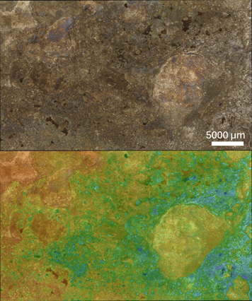
General material visualization tool. Feature depth and 3D visualization possible.
Scanning Electron Microscopy (SEM)
A scanning electron microscope (SEM) is an imaging technique that uses electrons to visualize the sample surface, the use of electrons results in a higher resolution. SEM can reach a resolution of more than 1 nanometer and the device operates in a high vacuum.

Energy-dispersive X-ray spectroscopy (EDX)
Considering the electron interaction volume in the sample caused by the primary electrons originating from the electron gun of the SEM, different types of interactions are present. Characteristic X-rays can be used in energy dispersive X-ray spectroscopy to detect and characterize the different elements present in the sample. According to Moseley’s law, each element has its own characteristic X-ray wavelength.
Electron backscatter detection (EBSD)
Electron Backscattered Diffraction (EBSD) is a Scanning Electron Microscopy (SEM) based technique. It allows for microstructure quantification, the measurement of texture, grain and phase boundary characterization, phase identification and strain determination in crystalline multiphase materials of any crystal structure. The use of this technique allows a better understanding of the mechanisms responsible for the fracture of materials.
Transmission Electron Microscopy (TEM)
In the transition electron microscope or TEM, a beam of electrons is transmitted through an ultrathin sample. While passing through it, the beam interacts with the sample whereby an image is formed. This image is magnified and focused onto an imaging device such as a fluorescent screen, or detected by a sensor such as a CCD camera. Since the TEM uses electrons as source instead of light, a resolution is reached which is thousand times better than with an optical microscope.
X-ray diffraction (XRD)
X-ray diffraction is used to investigate the atomic arrangement of a crystal. An atom can scatter X-rays in all directions, but when atoms are periodically arranged, i.e. in a crystal, there are certain directions and angles in which the outgoing X-rays have a constructive interference. This occurs when Bragg’s law is satisfied. Since different phase have a different atomic arrangement, it is possible to distinguish several phases with XRD in a heterogeneous compound.
Grazing incidence XRD (GIXRD) allows for better determination of crystallographic phases at the sample surface by fixing the source at low angles, limiting the interaction volume to only the top surface layer.
Spark source optical emission spectroscopy (SS-OES)
This is an quick technique to perform a quantitative metal analysis. It has a high sensitivity and reproducibility and has excellent detection levels for carbon, nitrogen and oxygen.