Visualizing static muscle morphology and dynamic muscle behaviour using ultrasound imaging and EMG
Target audience
PhD and post-doc researchers with an interest in, and basic knowledge of US, gait analysis and EMG.
Organizing and scientific committee
Drs. Babette Mooijekind
Prof. Dr. Lynn Bar-On
Abstract
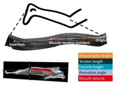
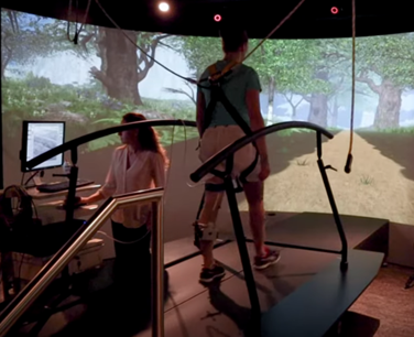
The application of ultrasound imaging (US) in musculoskeletal disorders has evolved with emergence of 3DUS, dynamic US, and integration of electromyography (EMG). With 3DUS it is possible to create a 3D reconstruction of a muscle. With this 3D reconstruction muscle morphological variables like muscle volume, muscle belly length, tendon length and muscle fascicle length can be calculated (Figure 1). With dynamic US we can visualize muscle behavior during dynamic conditions, such as walking and running (Figure 2). These techniques together can provide fundamental insight into pathology and the working mechanisms of treatments. In this course, PhD-students with interest in musculoskeletal imaging and/or EMG expand their knowledge by attending lectures and demonstrations by international experts in the field.


Objectives
This two-day course will start with lectures discussing the applications of musculoskeletal imaging (3DUS and dynamic US (Cenni et al., 2020; Cenni et al., 2018)), extracting relevant muscle variables, and several EMG modalities including high-density EMG and bipolar US-transparent EMG (Botter et al., 2019). After the students gained insight into the theory behind musculoskeletal imaging and EMG, demonstrations will be given by Dr. Cenni collecting and processing 3DUS and dynamic US data (day 1), and by Prof. Botter and Elena Cesti demonstrating high-density EMG and integrating dynamic US with EMG (day 2). The students can obtain hands-on experience collecting imaging data, and thereby develop the skills to accurately perform these measurements. Additionally, the students will learn to distinguish between good and bad quality signals.
Dates and Venue
21 and 22 February 2024, B3 UZ Gent and SMART SPACE UZ Gent.
Programme
Day 1: 21 February 2024
- 9h00 to 13h00: Dr. Francesco Cenni
- Theoretical principles of 3DUS for the assessment of muscle morphology.
- Hardware and software for 3DUS for the assessment of muscle morphology.
- Demonstration of 3DUS for the assessment and the extraction of morphological parameters with focus on the medial gastrocnemius and rectus femoris.
- 13h30 to 17h30: Dr. Francesco Cenni - dynamic US
- Theoretical principles of dynamic US imaging.
- Semi-automatic tracking of the medial gastrocnemius muscle-tendon junction during gait.
- Demonstration of dynamic US imaging of the medial gastrocnemius during gait.
Day 2: 22 February 2024
- 09h00 to 13h30: Prof. Alberto Botter and Elena Cesti
- The principles and applications of high-density EMG
- Integration of EMG with ultrasound.
- 14h00 to 16h30: Group exercises
Registration
Follow this link for the registration and waiting list.
Registration fee
Free of charge for Doctoral School members.
The no show policy applies.
Number of participants
Maximum 13
Language
English
Evaluation method
100% attendance
After successful participation, the Doctoral Schools will add this course to your curriculum of the Doctoral Training Programme in Oasis. Please note that this can take up to one to two months after completion of the course.