Multiscale modelling of lymph transport
Introduction
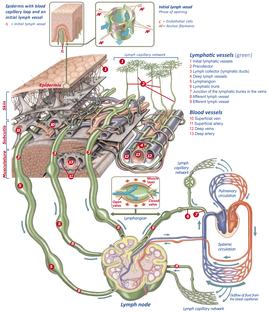
The lymphatic system is a tissue drainage network of converging vessels that maintains the tissue homeostasis by transporting the excess fluid, proteins, and metabolic waste products from the interstitium and ultimately returning it to the venous circulation. Impairment of lymph propulsion can lead to various pathologies including lymphedema which manifests itself by excessive swelling of the extremities. Currently, there is no long-term treatment available for lymphedema and its management aims at reducing or delaying progression of the swelling. Lack of a proper understanding of the fluid transport mechanisms in the lymphatic system has in part led to the absence of effective treatments. The aim of our research is to gain fundamental insight into the determinant factors of mass transport in the lymphatic micro- and macro-circulation via mathematical modelling as well as ex-vivo experiments in rodents.
Collecting fundamental data on the anatomy of the lymphatic system in mice
In order to collect morphological information of the lymphatic system, we’ve been performing two different sets of animal experiments. The goal of the in vitro experiments is to collect morphological information at the macro-level to capture the bigger collecting lymphatic vessels in the fore limbs and hind limbs of the mice. In order to visualize the lymphatic vessels in the micro-CT scanner, contrast agent is interstitially injected into the lymphatic vessels. The produced images are then segmented and 3D reconstructed, resulting in the 3D representations shown in Fig. 2.
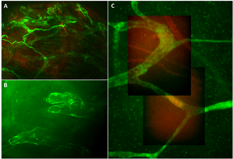
As a second approach, a clearing technique is used to collect morphological data at the micro-scale of the network of initial lymphatics with diameters of 10 µm or less. These sets of experiments require clearing and labelling of skin segments taken from mice limbs. Using the clearing technique, we are no longer constrained by the resolution limitations of micro-CT nor need to incorporate contrast/dye injections. The clearing protocol is performed in collaboration with the University of Antwerp. The cleared skin samples are then imaged and further processed (Fig. 3).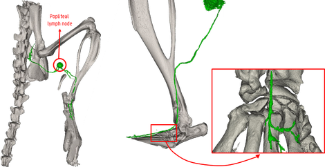
Multiscale computational modelling of lymph transport
Macro-scale (Lymph propulsion)
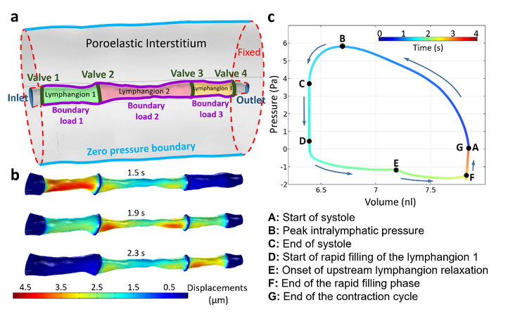
Collecting lymphatics are comprised of easily identifiable segments, called lymphangions, which are separated by secondary lymphatic valves. Lymphangions are the contractile components of the lymphatic system that actively transport the lymph along the lymphatic system (i.e. lymphatic propulsion). In order for lymph to return to the venous circulation, it travels against an adverse pressure gradient and gravitational forces. Hence, the active contraction of lymphangions (intrinsic pumping) and the presence of unidirectional secondary valves are of utmost importance in this process. Another contributing factor to lymph propulsion is extrinsic (passive) pumping, the deformation of tissues surrounding lymphatic vessels as a result of muscle contractions, arterial pulsations, etc. We have developed computational models of lymph propulsion using COMSOL Multiphysics® to study the behavior of flow within the collecting vessel, the deforming vessel wall, and the poroelastic interstitium surrounding it. Furthermore, by performing a parametric study, we can show how different parameters can positively and/or negatively impact the propulsion of lymph in healthy and impaired scenarios. A 3D computational model of lymph propulsion based on the 3D reconstructed morphological data obtained from the micro-CT imaging data of a mouse’s hind limb, is presented in Fig. 4.
Micro-scale (Lymph uptake)
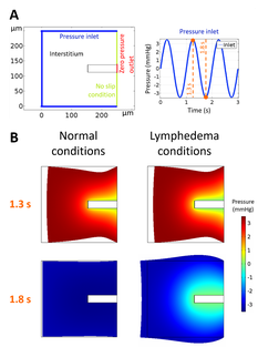
Terminal lymphatics are blunt ended permeable vessels made of a single layer of lymphatic endothelial cells. It is at the terminal lymphatics that the interstitial fluid enters the lymphatic system, in a process that can be called lymph uptake. The endothelial cell junctions act like unidirectional valves that allow lymph to enter inside the vessels while preventing lymph flow from returning to the interstitium. The changes in the interstitial pressures facilitate the opening of the primary lymphatic valves and lymph uptake. By developing a 2D computational model of lymph uptake in a single terminal lymphatic vessel, we aimed to mimic the function of primary lymphatics in both normal and lymphedema cases. We were able to show that lymphatic leakage through primary valves back into the poroelastic interstitium could drastically decrease the clearance rate of lymph leading to fluid accumulation and swelling of the surrounding tissue (Fig. 5). Our model can simulate different levels of impairment of lymphatic capillaries, from total obstruction of the primary valves to severely leaking ones.
Video material
In the following video, the 3D reconstructed model of lymph propulsion and its results are explained in a 3-minute pitch format.
IBiTech researchers currently active on the project
- Ghazal Adeli Koudehi (contact)
- Carlos Alejandro Silvera Delgado (contact)
- Patrick Segers (contact)
- Charlotte Debbaut (contact)
- Matthias Van Impe (contact)
Funding sources
- Flanders (FWO-Vlaanderen) – 3G022117- Multi-scale modelling of interstitial-to-lymphatic mass transport for the study of edema and trans-lymphatic drug administration (Patrick Segers, Charlotte Debbaut & Pieter Cornillie)
