Treatment planning of partial nephrectomy procedures
What is a partial nephrectomy?
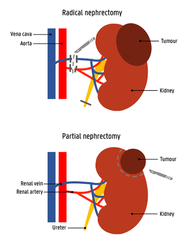
More than 90% of the renal cancers diagnosed in adults are renal cell carcinomas (RCC). When being diagnosed with an RCC, the first important step is to assess the stage of the tumour, as the entire treatment pathway will depend on this. In state I and II, the tumour is limited to the kidney and has not metastasized yet. Therefore, surgical removal of the tumour is the recommended treatment in these two first stages. Until recently, removing the entire kidney via a radical nephrectomy (RN) procedure guaranteed the best outcomes. Now, only the tumour is removed during a partial nephrectomy (PN) procedure. Both procedures are compared in Figure 1. PNs offer similar surgical morbidity and cancer control as RNs, while saving a large part of the kidney’s function. Therefore, a PN is preferred whenever possible [1]. However, during the execution of a PN, the surgeon faces several obstacles.
Before an incision can be made to remove the tumour, the blood supply to the tumour needs to be clamped off. Otherwise, the patient would lose too much blood and the vision of the surgeon would be impaired. Therefore, in the current practice, before the first incision is made, the main renal artery is clamped. In this way, the kidney and tumour are not perfused anymore and the tumour can be resected. The main problem with this approach is the limited time to execute the procedure. When the warm ischemic time (WIT) of the kidney exceeds 20 minutes, which means the blood supply to the kidney is interrupted for this amount of time, irreversible damage to the healthy tissue is occurring. This compromises the remaining functionality of the kidney [2]. To avoid this, the surgeon wants to clamp only the arteries that go to the tumour. This approach is called selective arterial clamping. As each kidney’s vasculature is different, the clamping positions need to be determined before surgery starts. Therefore, this research focuses on the development of a planning tool for partial nephrectomy procedures.

In the next video, this challenge is explained in one minute:
Development of a planning tool
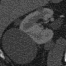
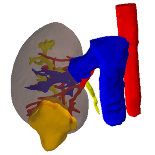
Patients diagnosed with an RCC usually get a CT-scan to assess the anatomy of the renal vasculature and the size and position of the tumour. This information, which consists of a stack of 2D-images as can be seen in Figure 2, is nowadays used to plan the clamping strategy. Using segmentation software, this 2D information can be segmented and 3D models can be generated, as shown in Figure 3. This results in a 3D visualisation of the kidney, the tumour, the blood supplying arteries and the relevant surrounding structures. Based on this information, surgeons can virtually test and optimize their approach using the planning tool software, e.g. giving estimations of the amount of viable and functional kidney tissue left after the procedure.
References
- M. C. Mir, I. Derweesh, F. Porpiglia, H. Zargar, A. Mottrie and R. Autorino. Partial Nephrectomy Versus Radical Nephrectomy for Clinical T1b and T2 Renal Tumors: A Systematic Review and Meta- analysis of Comparative Studies. European Urology, 71(4):606 – 617, 2017
- R. H. Thompson, et al. Every minute counts when the renal hilum is clamped during partial nephrectomy. European Urology 58 (3); 2010. 340 –345.
IBiTech researchers currently active on the project
- Charlotte Debbaut (contact)
- Karel Decaestecker (UZGent) (contact)
- Charles Van Praet (UZGent) (contact)
- Pieter De Backer (contact)
- Saar Vermijs (contact)
Funding sources
- BOF starting grant – grant BOFSTA201909015 2019-2023 (Charlotte Debbaut)
- Financial support by Ipsen (Karel Decaestecker)
- VLAIO Baekeland mandate HBC.2020.2252 (Pieter De Backer)


