Soft tissue characterization
Shear wave elastography

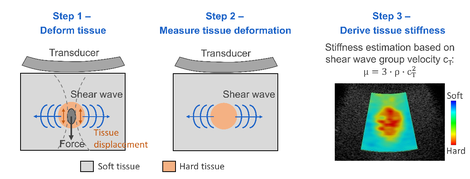
SWE has yet proven its clinical added value in several domains: differentiating malignant (stiff) from benign (soft) breast lesions [6] or staging liver fibrosis [7]. However, SWE could also advance clinical diagnosis for other tissue applications, where the tissue material characteristics include anisotropy, viscoelasticity and nonlinearity. Preliminary studies in literature have shown that shear wave physics in these settings are far more complex, implying that previously developed imaging setups and signal processing cannot simply be transferred to these applications. There is thus a clear need to address fundamental research questions reconciling the ultrasound physics with the complex cardiovascular biomechanics. Therefore, our lab studies these research questions using simulations, experiments or a combination of both. We are also closely collaborating with ultrasound labs at the Cardiovascular Imaging and Dynamics lab (KULeuven, Leuven, Belgium),Thorax center BME (Erasmus MC,Rotterdam, the Netherlands) and Nightingale lab (Duke University, Durham, NC, USA) to directly translate and potentially implement our findings to their experimental ultrasound systems.
Multiphysics approach for shear wave elastography
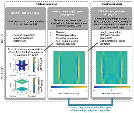
Our modeling framework for SWE combines biomechanical simulations of shear wave modeling with a simulation model of US imaging [8-9], as shown in Fig. 2. Specifically for SWE-modeling, shear waves are excited by applying a temporal and spatial varying body force to our finite element model, which can be derived from acoustic pressures simulated in Field II. The Field II settings can be derived from complementary SWE experiments, which we aim to mimic. Then, the actual SW propagation is modeled with the Finite Element software Abaqus, in which the material model is ideally calibrated with mechanical experiments. The second step in this multiphysics approach creates synthetic images of the SW propagation, mimicking the settings of our experimental set-up.
Using this complete approach or part of this approach, multiple areas of SWE have been studied:
- Understanding the effects of complex material properties on shear wave propagation patterns (including wave dispersion)
- Providing a 3D understanding of the shear wave field
- Studying the influence of pathology (increased tissue stiffness) on shear wave propagation characteristics
- Identifying correct material models based on experimentally observed shear wave propagation patterns
- Investigating the performance of ultrasonically tracking the shear wave displacement field
Advancing soft tissue characterization
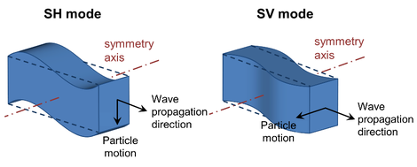
Current SWE measurements have mainly focused on the excitation and analysis of one specific shear mode, i.e. the shear horizontal mode (SH). For this mode, the wave polarization vector is perpendicular to the plane formed by the material symmetry axis and wave propagation direction (see Fig. 3). However, it is theoretically known that analysis of additional wave modes such as the shear vertical mode (SV) – defined by a wave polarization vector parallel to the plane formed by the material symmetry axis and wave propagation direction – can be used to advance material characterization for soft tissues with complex material properties.

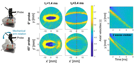
In collaboration with the Nightingale lab at Duke university, we have shown proof-of-concept in an uniaxially stretched phantom that it is experimentally possible to induce both shear wave modes at the same time by tilting the excitation axis 45⁰ with respect to the material’s symmetry axis, as visualized in Fig. 4. Complementary finite element simulations were performed using material parameters determined from mechanical testing, which enabled us to convert the observed shear wave behaviour into a correct representative constitutive law for the phantom material, i.e. the Isihara model. More information about this work can be found in [10-12].
Based on these findings, the Nightingale lab at Duke University is currently investigating whether multiple shear wave modes can also be excited in an in vivo setting.
References
-
H. Kanai, et al., Propagation of spontaneously actuated pulsive vibration in human heart wall and in vivo viscoelasticity estimation, IEEE TUFFC, 2005.
-
L. Gao, et al., Sonoelasticity imaging: theory and experimental verification, JASA, 1995.
-
A. P. Sarvazyan, et al., Shear wave elasticity imaging: a new ultrasonic technology of medical diagnostics, UMB, 1998.
-
K. Nightingale, et al., ARFI: in vivo demonstration of clinical feasibility, UMB, 2002.
-
J. Bercoff, et al., Supersonic shear imaging: a new technique for soft tissue elasticity mapping, IEEE TUFFC, 2004.
-
R. G. Barr, et al., WFUMB guidelines and recommendations for clinical use of ultrasound elastography. Part 2: breast, UMB, 2015.
-
G. Ferraioli, et al., Shear wave elastography for evaluation of liver fibrosis, J. Ultrasound Med., 2014.
- M.L. Palmeri, et al., A finite-element method model of soft tissue response to impulsive acoustic radiation force, IEEE TUFFC, 2005.
- M.L. Palmeri, et al., Ultrasonic tracking of acoustic radiation force induced displacements in homogeneous media, IEEE TUFFC, 2005.
- A. Caenen, et al., Measuring nonlinearity in a soft solid using a tilted acoustic radiation force for shear wave excitation, IEEE IUS, 2019.
- N. C. Rouze, et al., Wave speeds and Green’s tensors for shear wave propagation in incompressible, hyperelastic materials with uniaxial stretch, IEEE IUS, 2019.
- A. Caenen, et al., Analysis of multiple shear wave modes in a nonlinear soft solid: experiments and finite element simulations with a tilted acoustic radiation force, JMBBM, 2020.
IBiTech researchers currently active on the project
Funding sources
Junior post-doctoral fellowship by Research Foundation Flanders FWO – grant 1211620N 2019-2022 (Annette Caenen)
PhD fellowship by Flanders Innovation and Entrepreneurship Agency VLAIO – grant 141010 2014-2019 (Annette Caenen)
Post-doctoral fellowship by Research Foundation Flanders FWO – grant 1276416N 2014-2016 (Abigail Swillens)
PhD fellowship by Research Foundation Flanders FWO – grant 11U2116N 2013-2018 (Darya Shcherbakova)
Finalized PhDs within IBiTech
A Biomechanical Analysis of Shear Wave Elastography in Pediatric Heart Models (Annette Caenen)
Relevant link


