FT-IMAGER
FT-IMAGER is a wide-range multi-purpose Fourier-Transform Infrared facility for Materials, Archaeological and Geological Research at Ghent University.
Setup
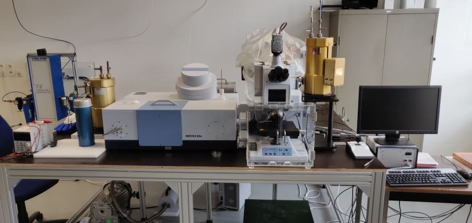
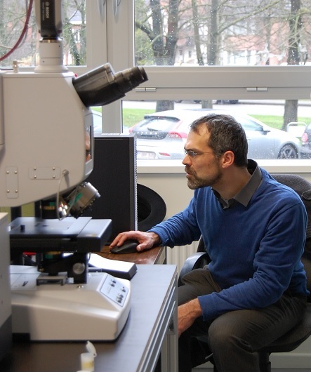
The setup is a combination of a vacuum near to far infrared spectrometer (Bruker Vertex 80v) and an FTIR microscope (Bruker Hyperion 2000).
It features a very large spectral range (10-15000 cm-1) and high spectral resolution (0.08 cm-1).
VERTEX-80v spectrometer
Standard
Transmittance-absorbance spectra in vacuo, at room temperature, plane parallel samples
Peripheral equipment
Liquid He cryostats for Mid- and Far IR ranges
Goniometer for specular reflectance measurements
Ge and KRS-5 crystal for attenuated total reflectance measurements
Gas cell, liquid/gas cell, liquids cells
HYPERION-2000 microscope
Standard
Sample at room temperature and ambient pressure, N2 flushed sample cabinet, motorized sample positioning system, mapping possibilities
Consortium
The consortium behind FT-IMAGER consists of research groups in solid state and semiconductor physics, inorganic and metal-organic chemistry, photonics, palaeontology and archaeometry.
Promoters
- Henk Vrielinck
Defects in Semiconductors (DiSC)
Department of Solid State Sciences
Faculty of Sciences - Klaartje De Buysser
Sol-gel Centre for Research on Inorganic Powders and Thin films Synthesis (SCRIPTS)
Department of Inorganic and Physical Chemistry
Faculty of Sciences - Pascal Van Der Voort
Center for Ordered Materials, Organometallics & Catalysis (COMOC)
Department of Inorganic and Physical Chemistry
Faculty of Sciences - Stephen Louwye
Research Unit Palaeontology (RUP)
Department of Geology and Soil Science
Faculty of Sciences - Kenneth Mertens
Research Unit Palaeontology (RUP)
Currently at IFREMER
Station Ifremer de Concarneau - France - Günther Roelkens
Photonics Research Group
Department of Information Technology
Faculty of Engineering and Architecture - Johan Lauwaert
Liquid Crystals and Photonics (LCP)
Department of Electronics and Information Systems
Faculty of Engineering and Architecture - Peter Vandenabeele
Archaeometry and Natural Sciences
Department of Archaeology
Faculty of Arts and Philosophy
Research
The main objective of the FT-IMAGER facility is to enhance FTIR research possibilities of all groups involved in the project.
A selection of studied topics is listed below.
Detection and identification of shallow- and semi-shallow- level defects in semiconductors
Detection of dopant and impurity levels in semiconductors
Shallow donors and acceptors introduce energy levels close to the conduction band and valence band, respectively, of a semiconductor.
At temperatures below the carrier freeze-out, these shallow levels are occupied with an electron (donor in n-type) or a hole (acceptor in p-type semiconductor).
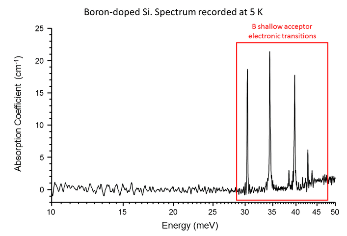
Then, in the far-infrared an additional absorption-lines-spectrum appears, characteristic for each shallow donor or acceptor.
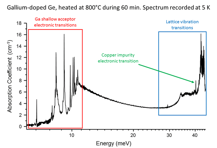
The figures below show the low temperature (<10K) far-infrared spectra of p-type silicon (B-doped) and germanium (Ga-doped). Note the logarthmic energy scale.
Typical ionization energies for shallow donors and acceptors in Si are 30-50 meV, and in Ge 9-15 meV.
The p-type Ge sample has received a high-temperature thermal treatment. This treatment introduces substitutional copper impurities in the lattice. Substitutional Cu introduces a semi-shallow energy acceptor (0/-) energy level in Ge at 40 meV. It is also visible in the far-infrared absorption spectrum.
This research is in part in collaboration with the Electro-Optical Materials division of Umicore.
Optical properties of absorber layers in thin-film solar cells
Thin-film solar cells offer the possibility of reducing material cost while maintaining an efficiency comparable to state-of-the-art Si solar cells.
Their absorber, in which light generates electron-hole pairs, is a very thin (a couple of µm) layer of a direct-band gap semiconductor, e.g. CdTe, CuIn(1-x)Ga(x)Se(2) (CIGS) or Cu(2)ZnSnS(4) (CZTS).
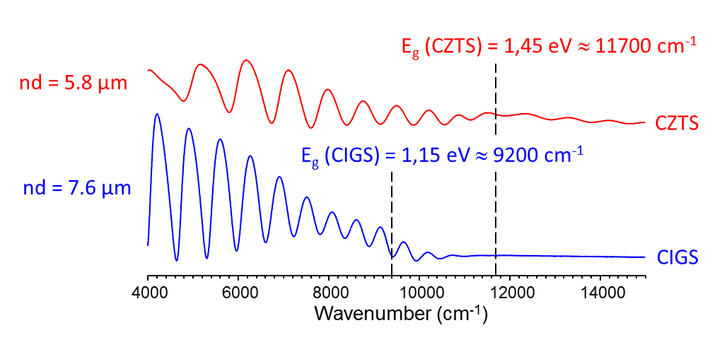
The figure below shows the near infrared reflectance spectra of a CIGS and a CZTS solar cell, measured with the FTIR microscope.
They exhibit a thin-film interference pattern, from which the optical thickness (product of refractive index n and thickness d) of the absorber layer (and other layers) in the cell can be determined.
For photon energies near and above the band-gap of the absorber layer, the interference franges get damped out due to absorption of light in the thin film.
At present Cu(8)SiSe(6) and Cu(2)ZnGeSe(4) absorbers layers on transparent and reflective substrates are investigated.
Their thickness and absorbance at photon energies below their band gap are deduced from micro-transmittance or -reflectance spectra.
These parameters are then used as input for numerical modeling (SCAPS) of solar cells based on these layers.
Part of this research is carried out in the framework of a European project (SWiNG) in which prof. Johan Lauwaert (LCP-group) is involved.
Vibrational mode study of Metal-Organic Frameworks in Mid- and Far-IR
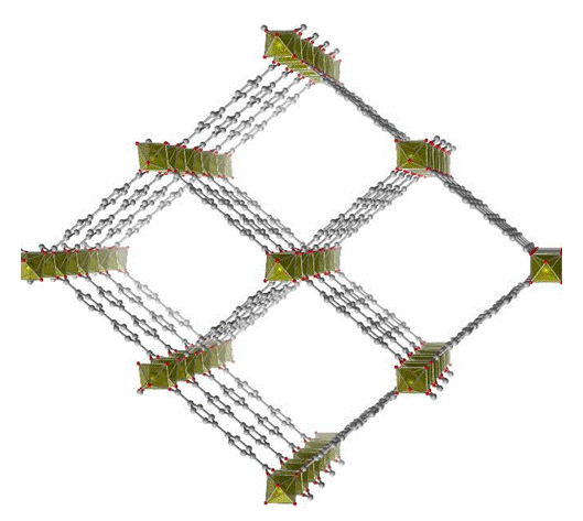
Metal-Organic Frameworks, or MOFs, are porous crystalline solids made up from metal-inorganic nodes, connetected by organic linkers.
MIL-53, or (M-OH)-benzene-dicarboxylate (M = Al, Ga, Sc, Cr, Fe) is prototypical example of a MOF, which exhibits a winerack-type topology.
Within this family of MOFs, a few exhibit breathing: they can undergo phase transitions between a state with narrow, closed pores and a state with large, open pores. The figure shows the structure for the large pore state. These transitions are reversible and are triggered by external factors, like temperature, pressure and the presence of certain guest molecules in the pores.
Alexander Hoffman examined the vibrational spectra of MIL-53(Al) in these two states, both experimentally and via molecular modelling, and published his results in a journal paper.
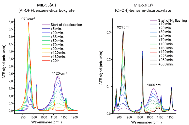
In the narrow pore state, MIL-53(Al) and MIL-53(Cr) reversiby adsorbe water in their pores. Hydration of the framework also has a marked influence on the vibrational spectra. Hence the hydration state of the framework can be followed in the infrared spectra.
The figure below shows the dehydration of MIL-53(Al) via desiccation of the air in the spectrometer sample compartment with silica gel (left), and of MIL-53(Cr) by flushing the sample compartment with dry nitrogen gas.
Upon dehydration, in both cases a broad band above 1050 cm-1 related to O-H bending gradually disappears and a sharper band below 1000 cm-1 grows in.
This research results from a collaboration between the research groups DiSC and COMOC with the Center of Molecular Modeling of UGent.
Contact
The FT-IMAGER facility is located at the Department of Solid State Sciences of Ghent University.
For more information about the facility, possibilities for collaboration and/or service measurements, please mail to Henk.Vrielinck@UGent.be.
Check out: FT-IMAGER in the VRT news
This facility is sponsored by the Hercules Foundation - FWO.
